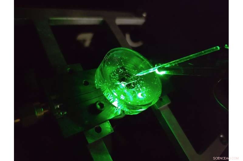
En prototyp av ett diamantspänningsmikroskop konstruerat av fysiker vid University of Melbourne. En liten elektrod är upphängd ovanför diamantchippet för att testa enhetens prestanda. En grön laser lyste underifrån ger fluorescensexcitation till chipet. Kredit:Författare tillhandahålls, University of Melbourne
Hjärnan är utan tvekan en av de mest komplexa strukturerna i det kända universum.
Fortsatta framsteg i vår förståelse av hjärnan och vår förmåga att effektivt behandla en mängd neurologiska sjukdomar är beroende av att undersöka hjärnans neurala mikrokretslopp med ständigt ökande detaljer.
En klass av metoder för att studera neurala kretsar kallas spänningsavbildning. Dessa tekniker gör att vi kan se spänningen som genereras av vår hjärnas avfyrande neuroner – berättar för oss hur nätverk av neuroner utvecklas, fungerar och förändras över tiden.
Idag utförs spänningsavbildning av odlade neuroner med hjälp av täta uppsättningar av elektroder på vilka celler odlas (eller odlas), eller genom att applicera ljusemitterande färgämnen som reagerar optiskt på förändringar i spänningen på cellens yta.
Men detaljnivån vi kan se med dessa tekniker är begränsad.
De minsta elektroderna kan inte på ett tillförlitligt sätt urskilja enskilda neuroner, cirka 20 miljondels meter i diameter, för att inte säga något om det täta nätverk av nanoskaliga förbindelser som bildas mellan dem, och inga betydande tekniska framsteg har gjorts på detta område på över två decennier.
Dessutom kräver varje elektrod sin egen trådbundna anslutning och förstärkare, vilket sätter betydande begränsningar för antalet elektroder som kan mätas samtidigt.
Färgämnen kan övervinna dessa begränsningar genom att avbilda spänningen trådlöst som ljus – detta innebär att den komplexa elektroniken kan placeras bort från cellerna i en kamera.
Resultatet är hög upplösning över stora ytor, som kan särskilja varje enskild neuron i ett stort nätverk. Men det finns begränsningar även här, spänningssvaren för toppmoderna färgämnen är långsamma och instabila.
Vår senaste forskning publicerad i Nature Photonics , utforskar en ny typ av en höghastighets, hög upplösning och skalbar spänningsbildplattform skapad i syfte att övervinna dessa begränsningar – ett diamantspänningsavbildningsmikroskop.
Utvecklad av ett team av fysiker från University of Melbourne och RMIT University, använder enheten en diamantbaserad sensor som omvandlar spänningssignaler på sin yta direkt till optiska signaler – detta betyder att vi kan se elektrisk aktivitet när den sker.
The conversion uses the properties of an atom-scale defect in the diamond's crystal structure known as the nitrogen-vacancy (NV).
NV defects can be engineered by bombarding the diamond with a nitrogen ion beam using a special type of particle accelerator. The fabrication of the sensor begins with using this process to create a high-density, ultra-thin layer of NV defects close to the diamond's surface.
You can think of each NV defect as a bucket that holds up to two electrons. When this bucket is empty, the NV defect is dark. With one electron, the NV defect emits orange light when illuminated by a laser—this property is known as fluorescence. With two electrons, the color of the fluorescence becomes red.
A previously discovered property of NV defects is that the number of electrons they hold—and the resulting fluorescence—can be controlled with a voltage. Unlike dyes, the voltage response of an NV defect is very fast and stable.
Our research aims to overcome the challenge of making this effect sensitive enough to image neuronal activity.
On the diamond's surface, the crystal structure ends with a layer one atom thick, made up of hydrogen and oxygen atoms. The NV defects closest to the surface are the most sensitive to changes in voltage outside the diamond, but they are also highly sensitive to the atomic makeup of the surface layer.
Too much hydrogen and the NVs are so dark that the optical signals we are looking for cannot be seen. Too little hydrogen and the NVs are so bright that the small signals we are after are completely washed out.
So, there's a "Goldilocks' zone" for voltage imaging, where the surface has just the right amount of hydrogen.
To reach this zone, our team developed an electrochemical method for removing hydrogen in a controlled way. By doing this, we've managed to achieve voltage sensitivities two orders of magnitude better than what has been previously reported.
We tested our sensor in salty water using a microscopic wire 10-times thinner than a human hair. By applying a current, the wire can produce a small cloud of charge in the water above the diamond. The formation and subsequent diffusion of this charge cloud produces small voltages at the diamond surface.
By capturing these voltages through a high-speed recording of the NV fluorescence, we can determine the speed, sensitivity and resolution of our diamond imaging chip.
We were able to further boost sensitivity by patterning the diamond's surface into 'nanopillars'—conical structures with the NV centers embedded in their tips. These pillars funnel the light emitted by the NVs towards the camera, dramatically increasing the amount of signal we can collect.
With the development of the diamond voltage imaging microscope for detecting neuronal activity, the next step is the recording of activity from cultured neurons in vitro—these are experiments on cells grown outside their normal biological context, otherwise known as test-tube or petri-dish experiments.
What differentiates this technology from existing state-of-the-art in vitro techniques is the combination of high spatial resolution (on the order of a millionth of a meter or less), large spatial scale (a few millimeters in each direction—which for a network of neurons in mammals is quite vast), and complete stability over time.
No other existing system can simultaneously offer these three qualities, and it's this combination that will allow our made-in-Melbourne technology to make a valuable contribution to the work of neuroscientists and neuropharmacologists globally.
Our system will aid these researchers in pursuing both fundamental knowledge and the next generation of treatments for neurological and neurodegenerative diseases. + Utforska vidare